Preliminary Program
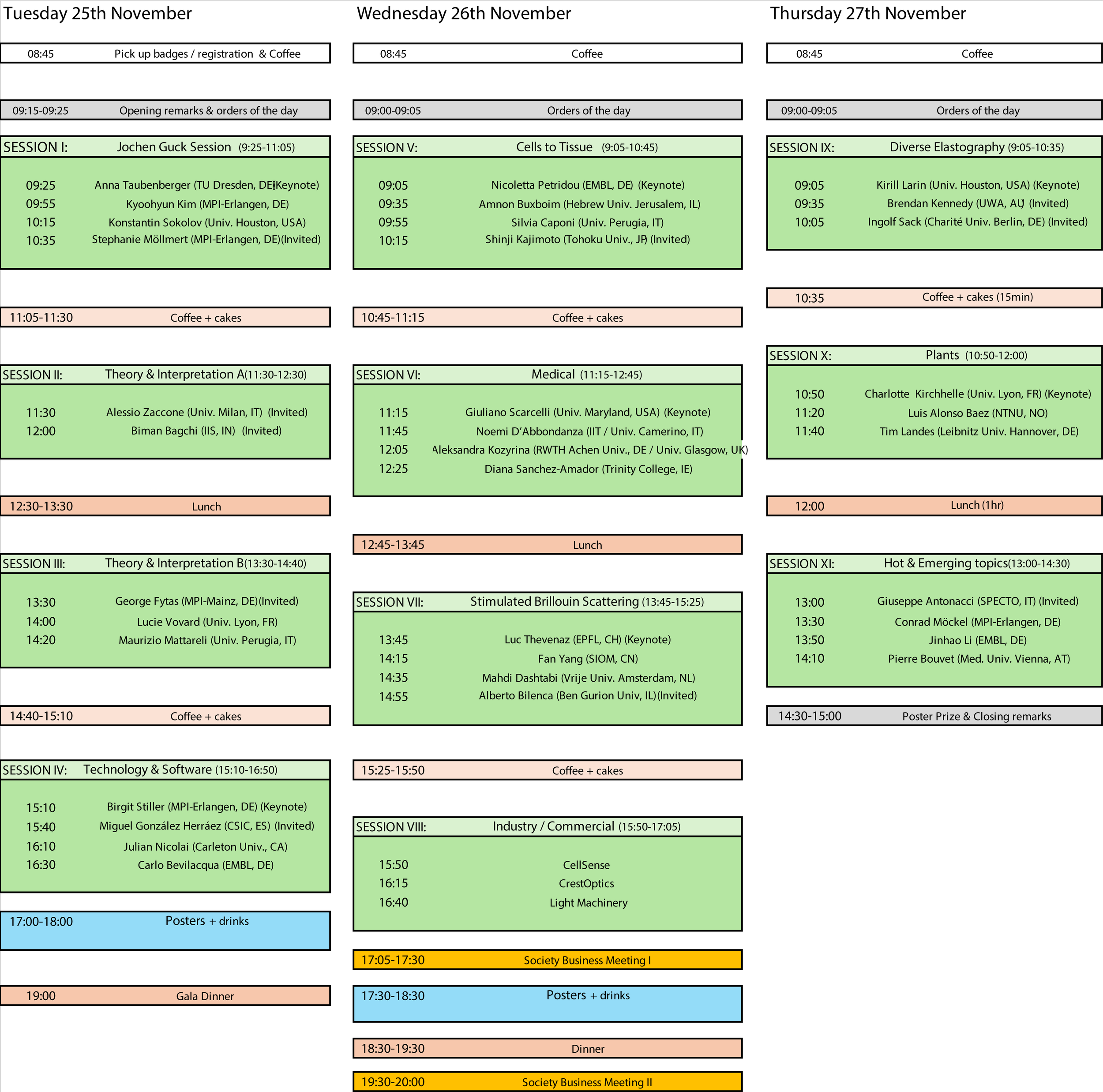
Talk titles
presenters, in chronological order of presentations
Session I: Jochen Guck Session
Anna Taubenberger (Keynote)
“Adaptations of cell biophysical properties during tumour growth and progression”
Kyoohyun Kim
“Optical Diffraction Tomography for quantifying cell physics”
Konstantin Sokolov
“Evaluation of changes in biomechanics of pancreatic cancer in response to therapy using Brillouin imaging”
Stephanie Möllmert (Invited)
“Mapping mechanical microenvironments in the female reproductive system”
Session II: Theory & Interpretation A
Alessio Zaccone (Invited)
“Bridging Brillouin scattering and mechanical testing of complex soft materials with atomistic modelling“
Biman Bagchi (Invited)
“Dynamics of Biological water“
Session III: Theory & Interpretation B
George Fytas (Invited)
“Brillouin Light spectroscopy applications in Soft Matter Science”
Lucie Vovart
“Network dynamics of compressed cell nuclei with Brillouin microscopy”
Maurizio Mattarelli
“Engineering elastic properties of Microparticles by charge irradiation”
Session IV: Technology & Software
Birgit Stiller (Keynote)
“Stimulated Brillouin scattering for photonic neuromorphic computing and quantum technologies”
González Herráez Miguel (Invited)
“Geophysical and biological applications of distributed optical fiber sensing in the oceans”
Julian Nicolai
"Ultra-Compact All-Etalon Cascaded Brillouin Spectrometer"
Carlo Bevilacqua
“A standardized file format and open-source analysis framework for Brillouin microscopy data”
Session V: Cells to Tissue
Nicoletta Petridou (Keynote)
“Tissue rigidity phase transition shapes morphogen gradients“
Amnon Buxboim
“Mechanical Characterization of Preimplantation Embryo Development Using Brillouin Microscopy”
Silvia Caponi
“Brillouin And Raman Micro-Spectroscopy For The Characterization Of Cells And Tissues”
Shinji Kajimoto (Invited)
“Quantification of intracellular LLPS using Raman and Brillouin microscopy”
Session VI: Medical
Giuliano Scarcelli (Keynote)
“Brillouin microscopy in vivo in the clinic”
Chiara Bartoli
“Brillouin microscopy on ALS-related protein aggregates in living cells”
Alesksandra Kozyrina
“The architecture of function: ECM-driven mechanical balance in retinal pigment epithelium”
Diana Sanchez-Amador
“Brillouin Microscopy analysis of macrophage viscoelasticity and polarisation in response to substrate stiffness and topography”
Session VII: Stimulated Brillouin Scattering
Luc Thevenaz (Keynote)
“Brillouin optical fibre sensing: principles and future perspectives”
Fan Yang
“Pulsed fiber laser system enables fast stimulated Brillouin scattering microscopy”
Mahdi Dashtabi
“Stimulated Brillouin Microscopy in the Telecom Band”
Alberto Bilenca (Invited)
“Progress and perspectives in stimulated Brillouin microscopy”
Session VIII: Industry/Commercial
CellSense
CrestOptics
LightMachinery
Session IX: Diverse Elastography
Kirill Larin (Keynote)
“Comparative Analysis of Several Elastography Techniques for Tissue Biomechanics“
Brendan Kennedy (Invited)
“Compression-based optical coherence elastography: mapping tissue elasticity on the micro-scale“
Ingolf Sack (Invited)
“Magnetic resonance elastography: cross-scale mechanical characterization of soft tissues from zebrafish to patients“
Session X: Plants
Charlotte Kirchhelle (Keynote)
“Cell edge polarity - a biophysical principle?”
Louis Alonso Baez
“Mapping Mechanical Properties in Diverse Tissues and Plant Species”
Timm Landes
“Brillouin- & Raman-scattering of tomato fruit cuticles”
Session XI: Hot & Emerging Topics
Giuseppe Antonacci (Invited)
“On-Chip Brillouin Spectrometer”
Conrad Möckel
“Optical Measurement of Mass Density of Biological Samples”
Jinhao Li
“Mass density and viscoelastic modulus measurement of biological samples enabled by all-optical correlative label-free imaging”
Pierre Bouvet
“Stimulation synchronized and co-localized angle-resolved Brillouin microspectroscopy for studying ultra-fast variations and 3D anisotropy of the Brillouin scattering spectra in biological samples”

Posters (Numbers, Titles & Presenters)
How big should my poster be?
Maximum A0 size. Please prepare your poster so that it fits on a vertical A0 sized poster wall.
Where and when should/can I hang up my poster?
Each poster is assigned a number (e.g. S1P1, S1P2, etc.). The poster walls in the Planck Lobby each have a number. Hang your poster on the poster wall that has the number of your poster. Please be sure your poster is up before 17:00 on the 25th November. You may use the time in the precluding coffee breaks or lunch break (or before meeting begin on the 25th November) to hang up your poster. Pins for mounting posters will be provided at the venue (ask at registration desk). Keep your poster up until the end of the second poster session (18:30, 26th November).
When should I be at my poster?
Depends…
November 25th 17:00-18:00 if the last digit of your poster number is odd (e.g. S1P1, S1P3, S2P1,…)
November 26th 17:30-18:30 if the last digit of your poster number is even (e.g. S1P2, S1P4, S2P2,…)
You are of course very welcome to be at your poster both days during the above times. (The purpose of the above divide is so that you can also have a chance to visit other posters and interact with their presenters).
Technology/software
S1P1 "Improving VIPA-Based Brillouin Spectrometers with Simulations and Electro-Optical Modulation"
B. Rufflé (Université de Montpellier, FR)
S1P2 “Toward Single-Shot Coarse 2D Brillouin Mapping Using a Multi-Pass Grating Spectrometer”
J. Nicolai (Carleton University, Ottawa, CA)
S1P3 “Electro-Optic Modulator source as sample-free calibrator and frequency stabilizer for Brillouin Microscopy”
C. Testi (Istituto Italiano di Tecnologia, Rome, IT)
S1P4 “Sample-free electro‑optic calibration for VIPA Brillouin microscopy”
M. Sng (Max Planck Institute / Friedrich-Alexander-Universität, Erlangen, DE)
S1P5 “Dual modality laser scanning confocal Brillouin and fluorescence microscopy for service in the EMBL Imaging Centre”
T. Ohn (European Molecular Biology Laboratory – Imaging Center, DE)
S1P6 “Non-Destructive Simultaneous Mapping of Cellular Stiffness and Ultrasmall Forces”
M. Harling (University of Cologne, DE)
S1P7 “Phonon microscopy for the characterisation of sperm cells“
F. Perez-Cota (University of Nottingham, UK)
S1P8 “Quantifying the effect of sample heterogeneity on Brillouin gain in SBS microscopy”
M. Xu (Shanghai Institute of Optics and Fine Mechanics, Shanghai, CN)
S1P9 “Universal Data Formats, analysis protocols, and visualization tools for Brillouin light scattering”
P. Bouvet (Medical University of Vienna, AT)
S1P10 “BrimView: an open-source Python web-app to visualize and analyze Brillouin microscopy data”
S. Hambura (European Molecular Biology Laboratory, DE)
S1P11 “Alternative Operation of Fabry–Perot Interferometers for Efficient 2D Brillouin Mapping”
M. Pochylski (Adam Mickiewicz University, PL)
Biophysics - Biology
S2P1 “Brillouin spectroscopy as a tool to quantify viscous response in soft matter”
Z. Wang (Heidelberg University, DE)
S2P2 “Exploring the protein-lipid interface of heterogeneous samples using light scattering spectroscopy”
S. Hill (University of Exeter, UK)
S2P3 “Multiscale Mapping of Oxidative Stress–Induced Mechanical Changes in the Central Nervous System”
J. Bachir Salvador (Max Planck Institute / Friedrich-Alexander-Universität, Erlangen, DE)
S2P4 “A role for microtubule order in squamous morphogenesis mechanobiology”
E. Huscavova (European Molecular Biology Laboratory, DE)
S2P5 “Cells decrease their volumes and cell cycle progression rates during formation of multicellular structures”
V. Mahajan (Technical University Dresden, DE)
S2P6 “From mental to mechanical stress: A Lamin B1 mediated story”
K. Hein (Max Planck Institute / Friedrich-Alexander-Universität, Erlangen, DE)
S2P7 “Phonon Microscopy Reveals Sub-Cellular Mechanical Adaptations in Rice Roots”
S. La Cavera (University of Nottingham, UK)
S2P8 “Biophysical methods in the field of parasitology”
A. Battistella (Max Planck Institute / Friedrich-Alexander-Universität, Erlangen, DE)
S2P9 “Spheroid mechanics in dynamic 3D microenvironments”
M. Valero Puigdomenech (Institute for Bioengineering of Catalonia, The Barcelona Institute for Science and Technology, ES)
Medical
S3P1 “Brillouin microscopy for the mechanical properties of vascular smooth muscle cells”
L. Zhang (Istituto Italiano di Tecnologia, Rome, IT)
S3P2 “Towards a biomechanical map of the human lung”
J.M. Gomez (European Molecular Biology Laboratory, DE)
S3P3 “Brillouin Microscopy Reveals Distinct Mechanical Signatures of White Blood Cells and mock Circulating Tumor Cells with potential for Liquid Biopsy application”
M. Behrouzitabar (Specto Slr, IT)
S3P4 “Bridging the gap in cancer mechanics with organoids”
M.G. Lettinga (Technical University Dresden, DE)
S3P5 “Blood plasma viscoelasticity: potential for post acute viral infection diagnosis”
J. Illibauer (Medical University of Vienna, AT)
S3P6 “Imaging the mechanical landscape of neurodegenerative disease”
M. Körbel (European Molecular Biology Laboratory, DE)
S3P7 “Assessing Mammalian Embryonic Tendon Mechanics using Brillouin Microscopy“
L. Sliney (Trinity College Dublin, IE)